Cervical Pillow for Dysautonomia and EDS — Why Cervical Support Matters Overnight
Sleep is generally treated as a passive state in dysautonomia management — something to optimize for duration and quality, but not a mechanically active intervention. That framing misses something important. The position of your body during sleep, and specifically the alignment of your cervical spine, is not neutral. It is actively read by the nervous system throughout the night, feeding continuous sensory information into the same regulatory circuits that govern autonomic output during waking hours. For people with hypermobility disorders and dysautonomia, the quality of that overnight sensory input is a variable worth managing.
The Upper Cervical Spine as an Autonomic Input
The upper cervical spine — the atlantoaxial and suboccipital region — is unusually dense with mechanoreceptors: sensory neurons that detect joint position, compression, and movement. These receptors feed directly into the brain's body map, contributing to the proprioceptive model the central nervous system uses to coordinate motor output, postural control, and autonomic regulation. The brainstem, which sits directly adjacent to the upper cervical region, is the hub of autonomic regulation: it is where vagal tone is modulated, where baroreceptor signals from the major vessels are processed, and where the coordination of heart rate, blood pressure, and respiratory rate is integrated.
When the upper cervical joints are in an abnormal position for an extended period, the mechanoreceptors in those joints send positional signals that do not accurately reflect the body's true state. The brain's regulatory output is calibrated, in part, on that incoming data. Inaccurate data produces inaccurate output. In a population where the autonomic system is already dysregulated, adding a source of inaccurate proprioceptive signaling — sustained across eight hours of sleep — is not a minor variable.
Hypermobility, Cervical Instability, and Autonomic Function
For people with hypermobile Ehlers-Danlos syndrome (hEDS) or hypermobility spectrum disorder (HSD), cervical instability is a common and underappreciated comorbidity. The same connective tissue laxity that affects joints throughout the body affects the ligamentous support structures of the cervical spine. Ligaments that are too compliant allow excessive movement at the atlantoaxial joint and the suboccipital segments — movement that the muscular system has to compensate for constantly, and that the proprioceptive system registers as chronic positional ambiguity.
Research on proprioceptive impairment in hypermobility and its link to autonomic dysfunction finds that the joint position sense of hypermobile individuals is systematically less accurate than in matched controls. The body map the autonomic system relies on for output calibration is built from proprioceptive input — and in hypermobility, that input is less reliable. Cervical instability amplifies this, because the upper cervical region is disproportionately important to the overall proprioceptive signal driving autonomic regulation.
The co-occurrence of hypermobility and dysautonomia is not coincidental. Hypermobility and dysautonomia share mechanisms that explain why they appear together so frequently — the same tissue and sensory abnormalities that produce joint laxity also interfere with the autonomic regulatory system's ability to receive and act on accurate body state information.
Head-of-Bed Elevation and Why Cervical Support Becomes Critical
Head-of-bed elevation — raising the head of the bed by 4 to 6 inches relative to the foot — is a documented intervention for POTS and orthostatic intolerance. The mechanism is primarily hormonal: sleeping at a slight incline maintains a mild orthostatic stimulus overnight, which stimulates the renin-aldosterone axis to retain more sodium and water. Over time, this tends to increase circulating blood volume without requiring any waking-hours effort. It is one of the few passive overnight interventions with a plausible mechanistic rationale for dysautonomia.
The practical challenge is that sleeping at an incline introduces a new positional variable: the cervical spine is no longer supported in the same geometry as flat sleep. A standard flat pillow maintains whatever geometry it was designed for on a horizontal surface. On an inclined surface, the head drops back or the neck flexes forward depending on pillow height and firmness, and either deviation from neutral creates the sustained positional stress described above.
A contoured orthopedic pillow designed to maintain neutral cervical alignment is not just about neck comfort. It is about ensuring that the proprioceptive input from the upper cervical region during elevated sleep is as accurate as possible — which, in a nervous system that is already struggling with regulatory calibration, is a meaningful intervention.
What to Look for in a Cervical Pillow for This Population
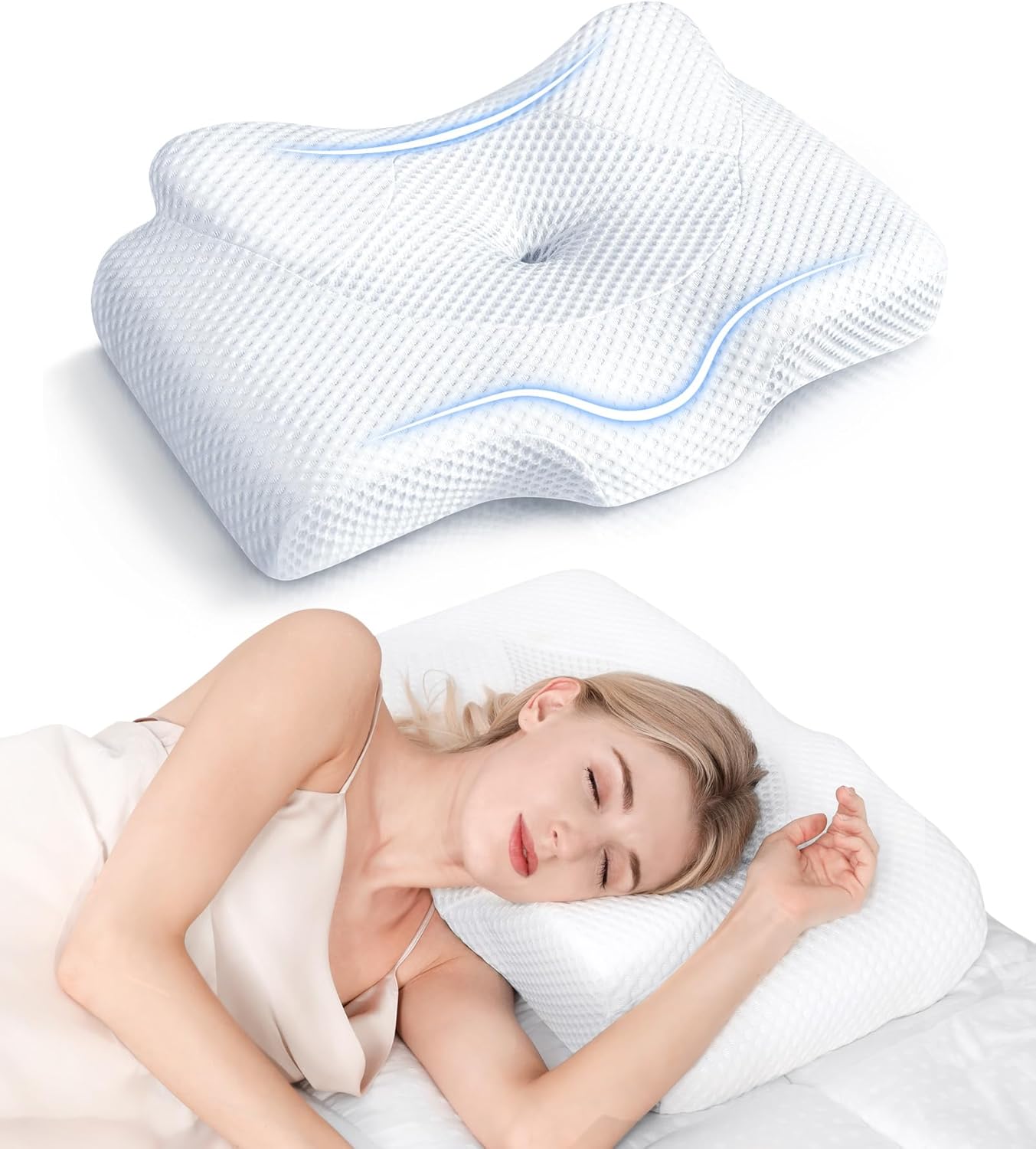
The critical feature is a contour that maintains neutral cervical alignment across position changes during sleep. Most people shift between back and side sleeping; the pillow needs to accommodate both without allowing the head to drop into hyperextension (too low) or cervical flexion (too high). Memory foam contour designs address this by providing differentiated zones — lower in the center for back sleeping, higher on the wings for side sleeping — so that the cervical curve is supported regardless of position.
The Osteo Cervical Pillow uses this contoured design with adjustable memory foam, which is particularly useful for a population where neck sensitivity is variable. Firm enough to actually support the cervical curve under body weight (critical for hypermobile joints that won't self-stabilize), but with enough give that it doesn't create pressure points at the occiput or shoulder. The goal overnight is eight hours of accurate cervical proprioceptive input — not comfort in the consumer sense, but appropriate support in the physiological sense.
Sleep position management is an underappreciated dimension of dysautonomia care. The cervical spine's connection to autonomic output is not hypothetical — it runs through the brainstem structures that sit directly adjacent to the upper cervical joints. Optimizing what those joints communicate to the brain throughout the night is a small intervention with potentially outsized downstream effects in a system that depends on accurate body-state information to function correctly.